


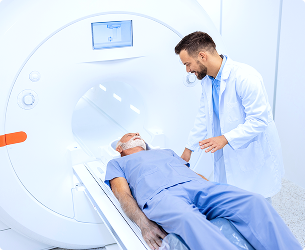
Image-Guided Biopsy
About the procedure
Care before the procedure
Thorough preparation ensures both safety and accuracy.
- Medical Evaluation: Review of medical history, current medications (especially blood thinners like warfarin, clopidogrel, or aspirin, which may need to be temporarily paused), and allergies (particularly to local anesthetics or contrast agents).
- Recent Imaging: Previous scans (e.g., MRI, CT, ultrasound) are reviewed to plan the best approach and access route.
- Pre-Procedure Instructions:
Fasting may be required for certain biopsies (e.g., abdominal biopsies) to reduce the risk of nausea if sedation is used. Patients should wear comfortable clothing and arrange for transportation if sedation is planned. - Informed Consent: The physician will explain the procedure, its benefits, potential risks (e.g., bleeding, infection, pain, rare injury to adjacent structures), and alternatives.
Care during the procedure
The procedure is typically quick, with continuous monitoring for patient comfort and safety.
- The Process: The patient is positioned to best access the biopsy site. The skin is cleaned and numbed with local anesthesia. Using real-time imaging, the physician inserts the biopsy needle through a tiny incision and advances it to the target. Multiple samples may be taken to ensure diagnostic adequacy.
- Patient Role: The patient must remain as still as possible and follow breathing instructions (e.g., hold breath briefly during needle advancement for chest/abdominal biopsies) to enhance precision.
- Sensations: Pressure or mild discomfort may be felt during needle insertion and sampling. Pain is generally minimal due to local anesthesia.
- Duration: Usually takes 30–60 minutes, depending on the location and complexity of the biopsy.
Care after the procedure
Post-procedure care focuses on monitoring, recovery, and follow-up.
- Immediate Aftercare: Pressure may be applied to the biopsy site to prevent bleeding. A small bandage is placed over the incision. Patients are monitored briefly for stability before discharge. Rest is advised for the remainder of the day; strenuous activity should be avoided for 24–48 hours.
- Symptoms to Watch For: Seek medical attention if there is significant swelling, redness, bleeding, fever, or worsening pain at the biopsy site.
- Results: Tissue samples are sent to a pathology lab for analysis. Results are typically available within 3–7 days and will be discussed by the referring physician in a follow-up appointment.
- Long-Term Management:
Results guide further treatment whether additional imaging, surgery, oncology referral, or continued monitoring. Normal results may require periodic follow-up imaging to ensure stability.
Malignant results will lead to a tailored treatment plan such as surgery, radiation, chemotherapy, or targeted therapy.
Relevant Specialties
Radiology
Radiology uses imaging to support the diagnosis, evaluation, and monitoring of a wide range of health conditions.
It plays a critical role in helping clinicians understand underlying medical concerns by providing clear and accurate visual insights, supporting informed decision-making across multiple specialties.
At KIMSHEALTH, radiology services are delivered through a patient-first approach, focusing on precision, efficiency, and timely support to ensure effective and coordinated patient care.
Oman
A trusted destination for specialised medical care supported by expert clinical professionals.
KIMSHEALTH Oman3
Facilities
80+
Doctors
30+
Specialties
20+
Services
Saudi Arabia
The largest multispeciality clinic in Jubail, offering quality medical care to the citizens.
KIMSHEALTH Saudi Arabia2
Facilities
50+
Doctors
20+
Specialties
10+
Services