


Magnetic Resonance Imaging (MRI)
About the procedure
Care before the procedure
Safety screening is the most critical step due to the powerful magnet.
- Comprehensive Screening: All patients must complete a detailed safety questionnaire. It is *ABSOLUTELY ESSENTIAL** to inform the
- Staff of any: Metallic implants (e.g., pacemakers, aneurysm clips, cochlear implants, neurostimulators – many are contraindications). Metal fragments in the body (e.g., from welding, shrapnel). Pregnancy or possibility of pregnancy.
- Contrast Agent: For some exams, a contrast agent (Gadolinium) is injected intravenously to enhance image detail. Inform staff of any kidney problems or allergies.
- Preparation: Instructions vary. For some abdominal MRIs, fasting for a few hours may be required. For others, no special preparation is needed.
- What to Wear: Patients will change into a gown without metal. All personal items (jewelry, watches, glasses, hairpins, hearing aids, and any clothing with metal zippers/threads) must be removed. Credit cards and electronic devices are prohibited as the magnet can erase/damage them.
Care during the procedure
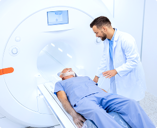
The process is painless but requires the patient to remain perfectly still.
- The Process: The patient lies on a motorized table that slides into the cylindrical opening of the MRI scanner. The technologist communicates via an intercom and can see the patient at all times.
- Patient Role: The most important task is to *remain as still as possible* throughout the scan, as movement blurs the images. The technologist may ask the patient to hold their breath for short periods during certain sequences.
- Sensations: The machine produces *loud, repetitive knocking, buzzing, and humming noises**. Earplugs or headphones are provided to protect hearing. Patients may feel a slight warming sensation in the area being scanned, which is normal. Some patients may feel anxious or claustrophobic inside the scanner; informing the team beforehand can lead to solutions like a mild sedative, an open MRI option, or a blindfold.
- Duration: The scan time typically ranges from *30 to 60 minutes*, depending on the body part being imaged.
Care after the procedure
There is typically no recovery time.
- Immediate Aftercare: If no sedation was used, patients can usually drive home and resume all normal activities immediately. If contrast was used, it is typically cleared by the kidneys within 24 hours.
- Results: The vast amount of data acquired requires detailed analysis by a radiologist. A formal report is sent to the referring physician, who will discuss the results with the patient, usually within a few days to a week.
- Long-Term Management: MRI is a cornerstone of modern diagnostic medicine. Its findings are used to make critical decisions about treatment strategies, surgical planning, and monitoring the effectiveness of therapy over time. Its lack of radiation makes it ideal for following chronic conditions that require repeated imaging. For patients with claustrophobia, discussing options like *open MRI* (less powerful magnet with open sides) or *wide-bore MRI** (a shorter, wider cylinder) with the referring doctor for future exams is advisable.
Relevant Specialties
Radiology
Radiology uses imaging to support the diagnosis, evaluation, and monitoring of a wide range of health conditions.
It plays a critical role in helping clinicians understand underlying medical concerns by providing clear and accurate visual insights, supporting informed decision-making across multiple specialties.
At KIMSHEALTH, radiology services are delivered through a patient-first approach, focusing on precision, efficiency, and timely support to ensure effective and coordinated patient care.

Oman
A trusted destination for specialised medical care supported by expert clinical professionals.
KIMSHEALTH Oman3
Facilities
80+
Doctors
30+
Specialties
20+
Services
Saudi Arabia
The largest multispeciality clinic in Jubail, offering quality medical care to the citizens.
KIMSHEALTH Saudi Arabia2
Facilities
50+
Doctors
20+
Specialties
10+
Services